Advanced Technology
Our Technology
At the office of Dr. Barry Rhome, we use the latest technology to diagnose, analyze and treat our patients. Our imaging systems are fast, safe, and efficient. These systems also integrate with our office software, which makes it easy to transfer and store any image for later reference. Our patients are our number one priority, and by using the latest technology, we aim to create a comprehensive and comfortable experience for you.
Digital Imaging
Barry Rhome DMD, PC uses state of the art Digital Imaging Technology to reduce the radiation exposure time by about 90% and provide detailed information that could be undetectable using traditional x-ray techniques.
Moreover, Dr. Rhome carefully chooses which and when radiographs are taken. There are many guidelines that we follow. Radiographs allow us to see everything we cannot see with our own eyes. Radiographs enable us to detect pathology in between your teeth, determine bone level, and analyze the health of your bone. We can also examine the roots and nerves of teeth, diagnose lesions such as cysts or tumors, as well as assess damage when trauma occurs.
Traditional dental radiographs are invaluable aids in diagnosing, treating, and maintaining dental health. Exposure time for traditional dental radiographs is minimal. However, Barry Rhome DMD, PC mainly utilizes Digital Imaging Technologies within the office. With digital imaging, exposure time is about 90% less when compared to traditional radiographs. Digital imaging can also help us retrieve valuable diagnostic information to see pathology.
Digital X-Rays
Dr. Rhome uses Digital X-rays to offer a more precise view of the tooth. The digital x-ray can be scaled and manipulated on the computer. These images can be stored and later retrieved to assess healing at a later check-up visit. Additionally, they can be easily and securely transferred via the computer to the patients’ other healthcare providers, if needed. Digital x-rays result in 1/6th radiation exposure to you, compared with traditional films.
Cone Beam CT

When a definitive diagnosis cannot be made with computerized digital x-rays, Dr. Rhome will utilize state-of-the-art, small volume cone-beam CT (computed tomography) technology that provides highly accurate, 3-D radiographic images. This allows three-dimensional visualization of teeth, bone, sinuses and surrounding structures with minimal radiation to the patient, enabling a level of anatomical accuracy and patient care not possible with 2-D technologies (regular dental x-rays or even digital x-rays). With the addition of cone-beam CT technology to our office, our practice is committed to providing innovative, high-quality, patient care.
Surgical Microscopes
The introduction of the surgical microscope has revolutionized the field of Endodontic Microsurgery. We have invested in the very best quality surgical microscopes, by Global Industries, that provide unparalleled magnification and illumination for our surgical procedures.
Our success depends on us being able to see the minutest of details—you cannot treat what you cannot see.
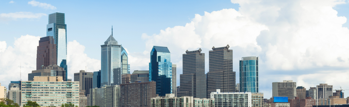
Contact Your Philadelphia Endodontist
Our technology enhances our expertise and ability to create personalized treatment plans for you and your family members. Dr. Rhome and his staff are happy to answer any questions you have about these imaging systems or anything else. To schedule your next appointment, contact Dr. Rhome at our Philadelphia office by giving us a call at 215-735-7113. We look forward to using these technologies to help serve you.